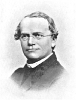
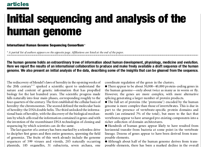
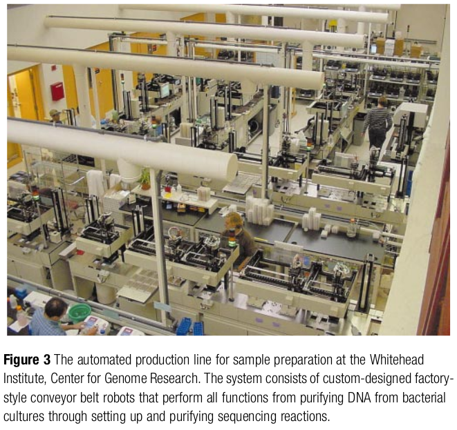
1866: Mendel’s paper on plant hybridisation was published.
1905: William Bateson used the term genetics in a letter.
1909: Wilhelm Johannsen introduced the term gene.
These terms preceded the identification of DNA as the principal hereditary material.
DNA as hereditary material
Avery, MacLeod, and McCarty identified DNA as the transforming principle in 1944.
Hershey and Chase provided further evidence using bacteriophages in 1952.
Watson and Crick proposed the DNA double helix in 1953, informed by key experimental work including that of Franklin, Gosling, Wilkins, and colleagues.
Chromosomes package long DNA molecules with associated proteins.
Familial hypercholesterolemia (FH)
…1972
M. S. Brown & J. L. Goldstein. Studied FH
They applied the concept of Inborn error of metabolism.
Postulated the existence of a genetic disease affecting the repression of the cholesterol synthesis.
Patients with FH showed large indexes of cholesterol in blood -> early cardiovascular troubles.
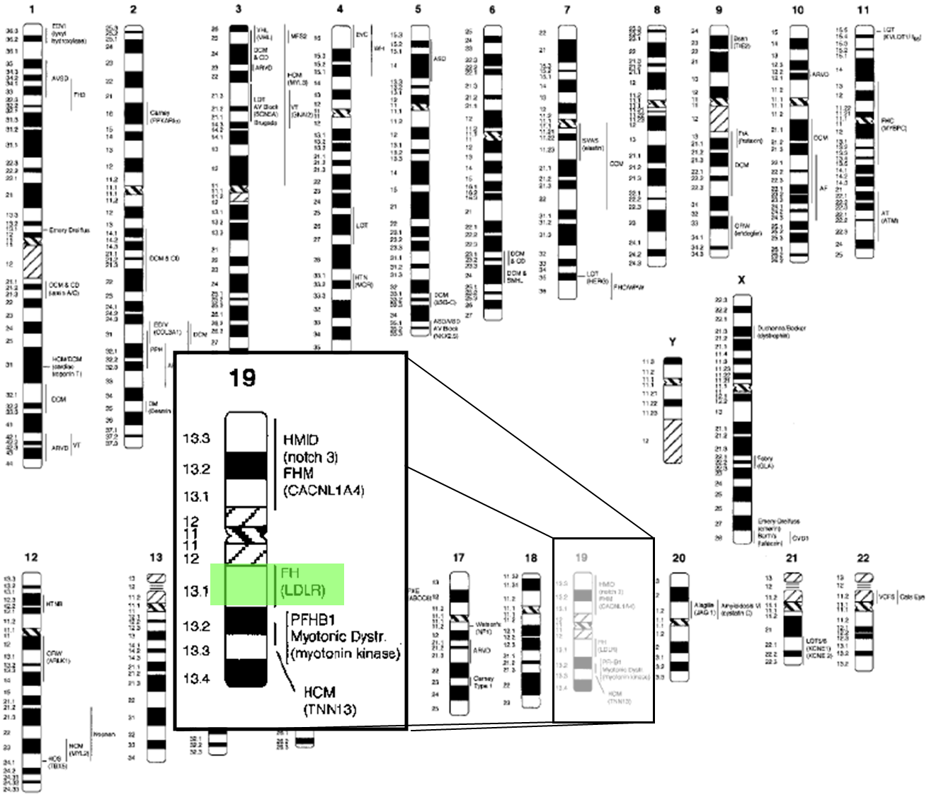
from Genetics of Cardiovascular Disease, Circulation, 102:20 (2000)
Cholesterol regulation in FH (1/2)
HMG-CoA reductase catalyses a rate-limiting step in cholesterol synthesis. HMG-CoA itself is a metabolite, not the enzyme.
Brown and Goldstein’s studies connected LDL uptake to regulation of cholesterol synthesis. Severe receptor defects disrupt this feedback.
Cholesterol regulation in FH (2/2)
The LDL receptor hypothesis
Brown and Goldstein linked defective LDL-receptor function to impaired cellular LDL uptake.
Their work explained how LDL uptake participates in feedback control of cholesterol synthesis.
Pathogenic variants can affect receptor production, transport, binding, internalisation, or recycling.
A genetic defect need not imply complete absence of the receptor.
Familial hypercholesterolemia
FH causes elevated LDL cholesterol and increases the risk of premature atherosclerotic cardiovascular disease.
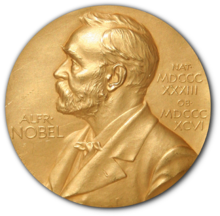
Brown and Goldstein received the 1985 Nobel Prize for discoveries concerning cholesterol metabolism.
Modern studies place heterozygous FH prevalence around 1 in 250–310, with variation by population and diagnostic criteria.
Individual risk depends on genotype, exposure over time, treatment, and other risk factors.
Genetic mechanisms and annotation
FH is a classic example of a disorder that can result from a pathogenic variant in a single gene.
Different genes can produce an FH phenotype, including LDLR, APOB, and PCSK9.
Connecting a variant to a phenotype requires evidence about the sequence, gene, molecular mechanism, and clinical context.
Recording these connections is a central purpose of biological annotation.
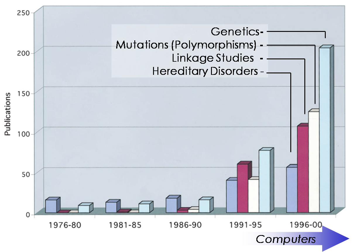
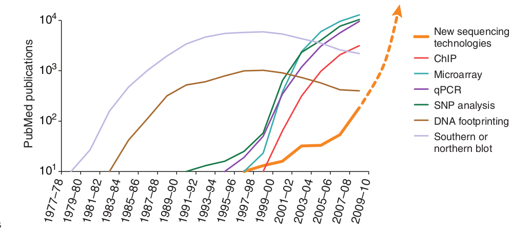
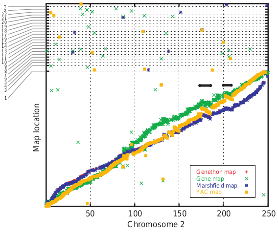
Evolution of publications in 25 years
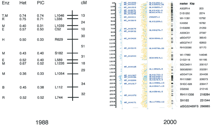
Chromosome 1 annotation: 1988 and 2000
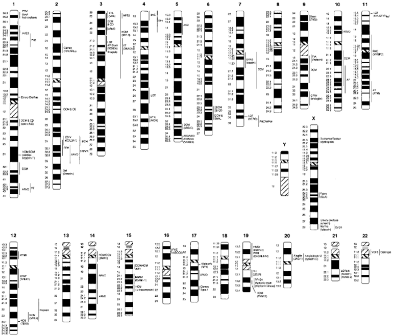
Evolution on (cardiovascular) annotations on Genome
Evolution on (cardiovascular) annotations on Genome
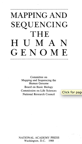
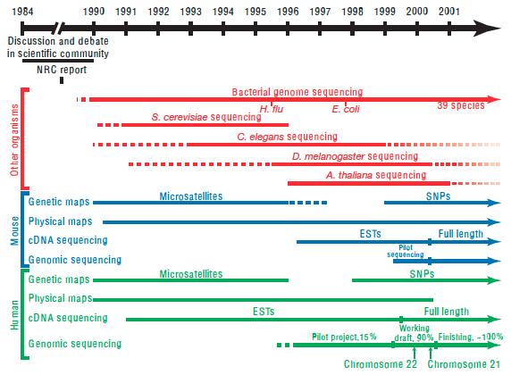
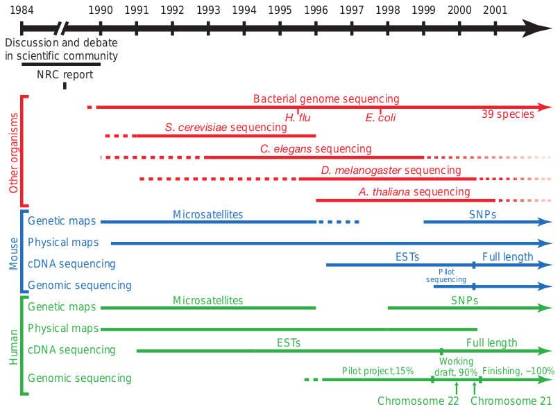
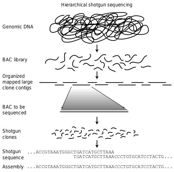
Human Genome Project?
Human Genome Project?
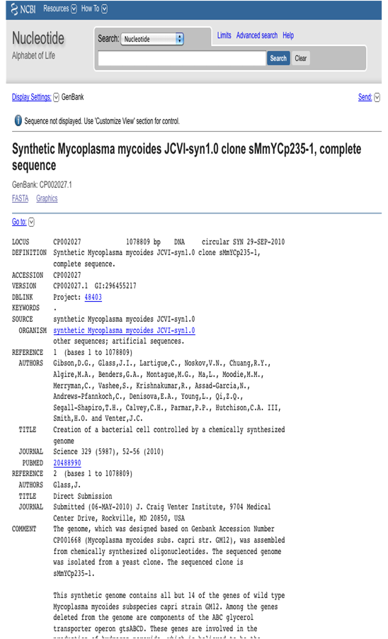
Craig Venter unveils “synthetic life”
A synthetic genome was transplanted into an existing recipient cell; the entire cell was not built from scratch.
TED · 18:18 · TED, 2010 · English subtitles from the published caption track.
Where are we now?
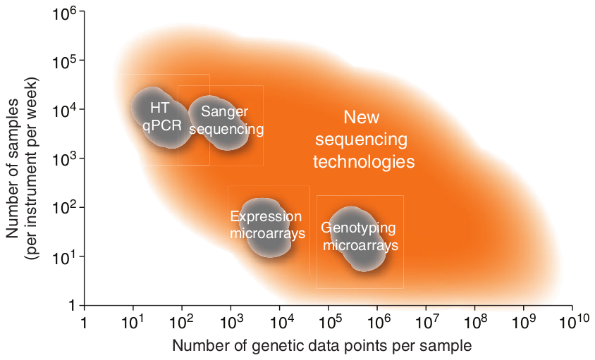
Evolution in genotypic technologies
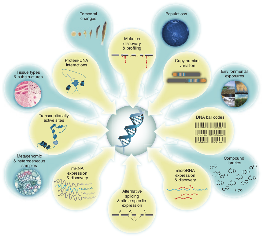
Kahvejian, A., Quackenbush, J., & Thompson, J. F. (2008). What would you do if you could sequence everything?. Nature biotechnology, 26(10), 1125-1133.
Evolution in genotypic technologies
Evolution in genotypic technologies
Evolution in genotypic technologies
DNA, RNA, and proteins
DNA
Stores hereditary information in a nucleotide sequence.
RNA
Participates in information transfer, regulation, catalysis, and cellular structure.
Proteins
Perform catalytic, structural, transport, and signalling functions. Protein sequences use 20 standard amino acids, with specialised exceptions.
DNA nucleotides
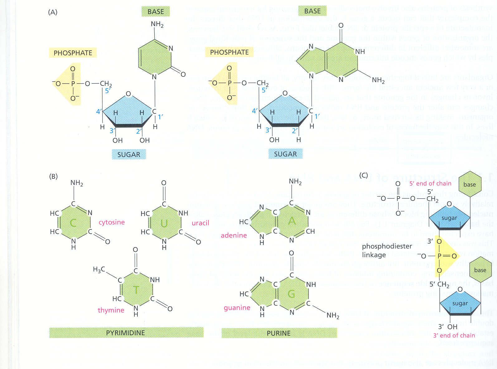
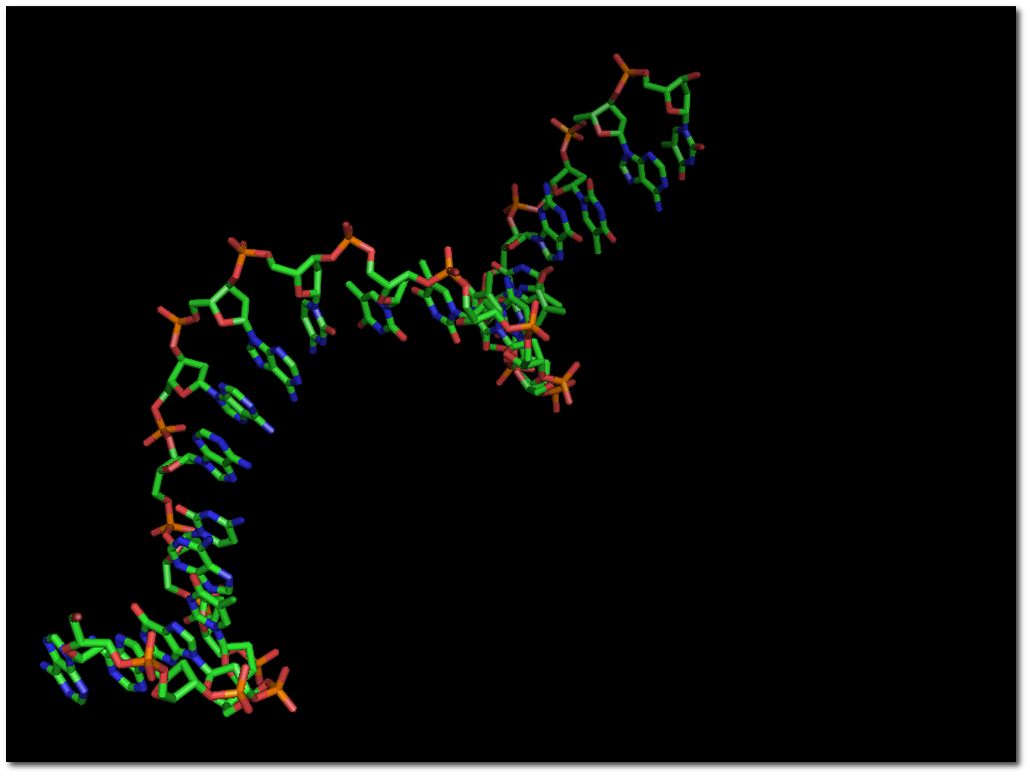
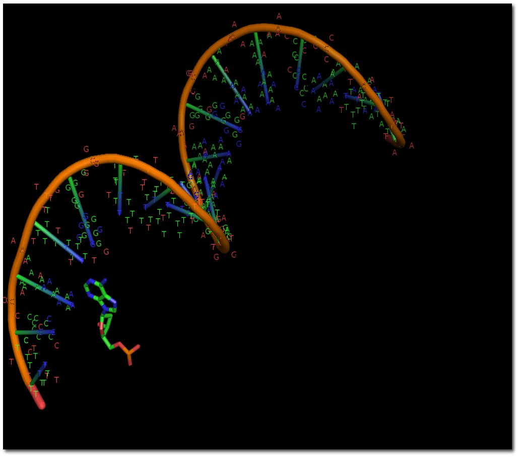
A DNA nucleotide contains a nitrogenous base, deoxyribose sugar, and phosphate.
Base class
DNA bases
Purines
Adenine (A), guanine (G)
Pyrimidines
Cytosine (C), thymine (T)
RNA contains uracil (U) in place of thymine. A haploid human nuclear genome contains roughly 3 billion base pairs.
DNA structure
DNA structure
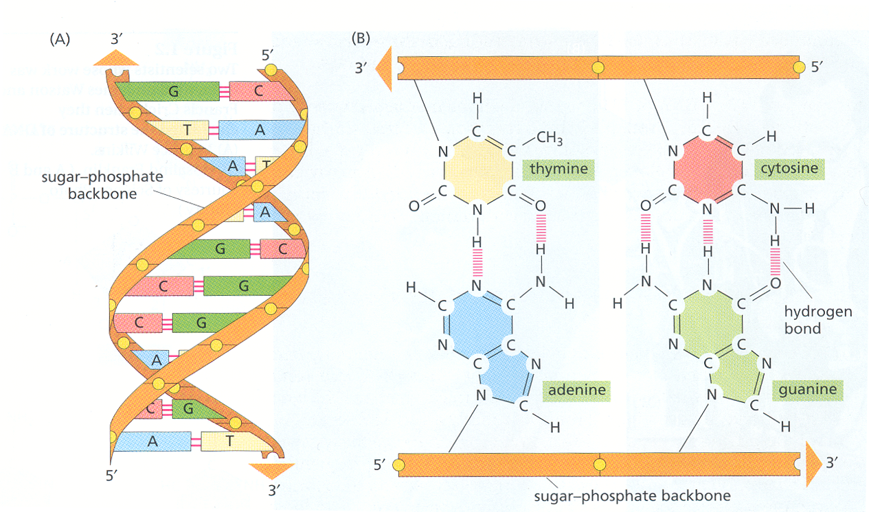
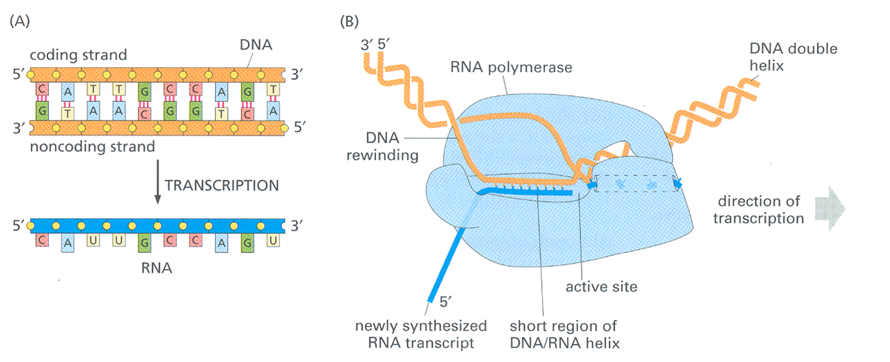
The resulting chain has a 5’ end (free phosphate group) and a 3’ end (free hydroxyl group).
The base sequence is defined as the order of the nucleotides along the chain from the 5’ to the 3’ end.
DNA structure
DNA replication
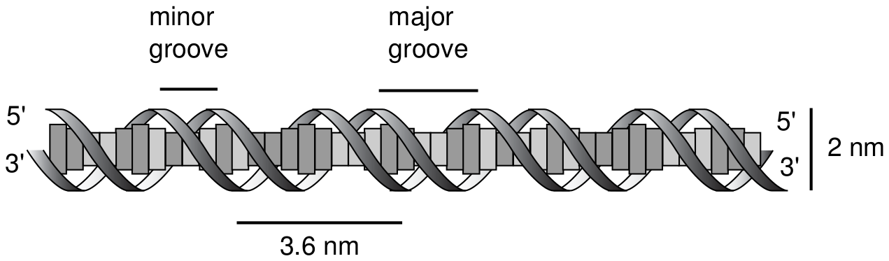
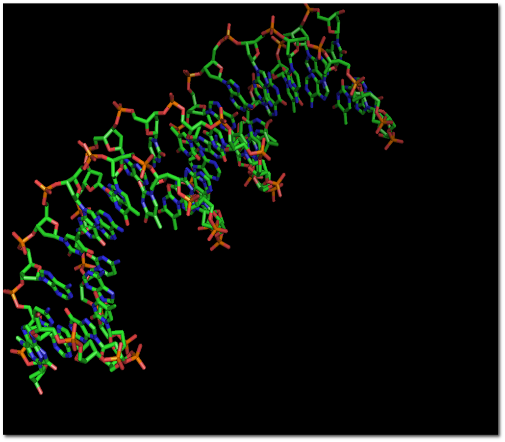
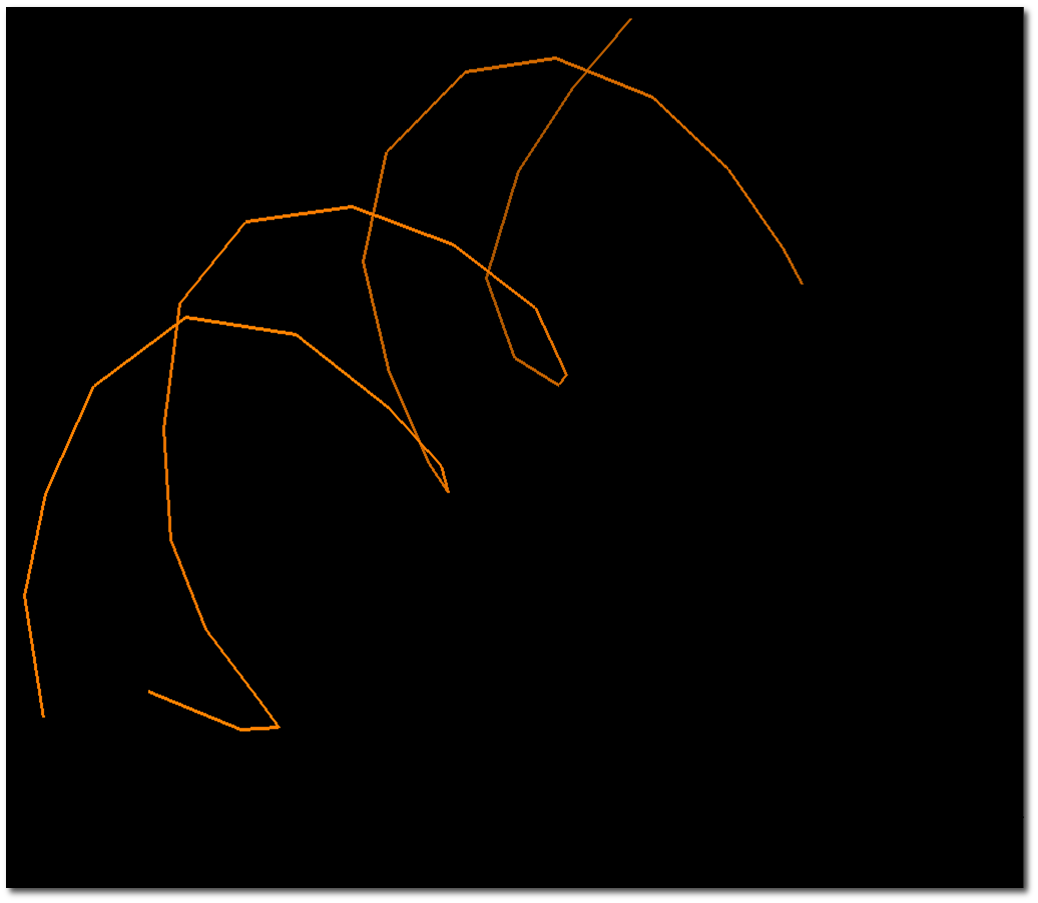
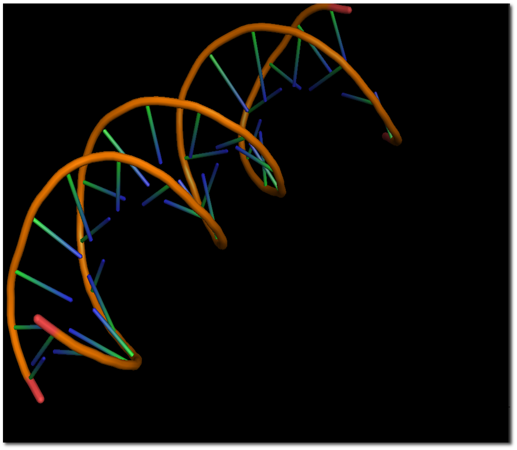
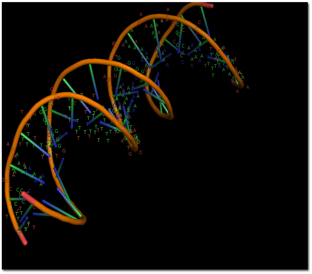
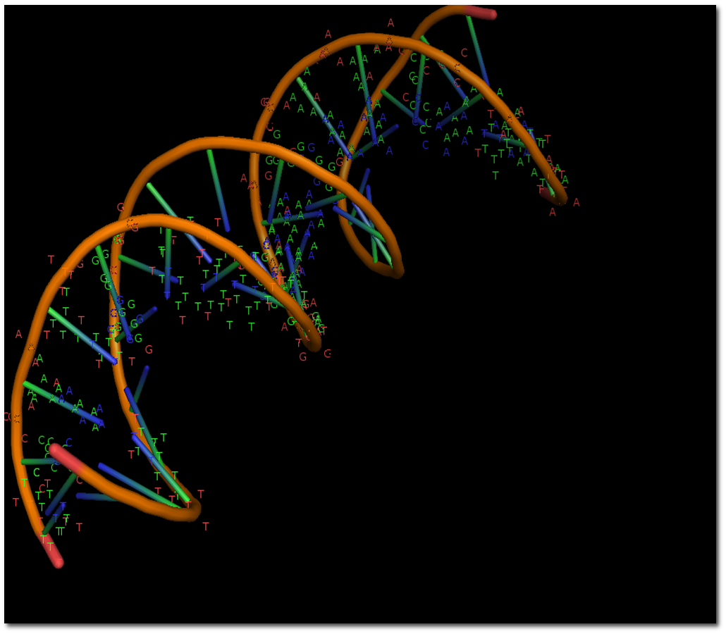
Complementary strands pair through hydrogen bonds; base stacking also stabilises the double helix.
The sugar–phosphate backbones lie on the outside of the helix.
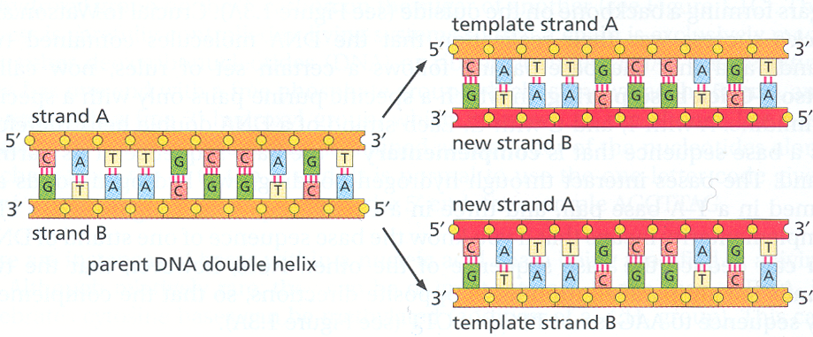
Polymerases synthesise DNA in the 5′ to 3′ direction using a template strand.
Proofreading and repair improve fidelity. Error rates depend on the organism and on which stage of replication or repair is measured.
Complementary DNA and RNA sequences can hybridise.
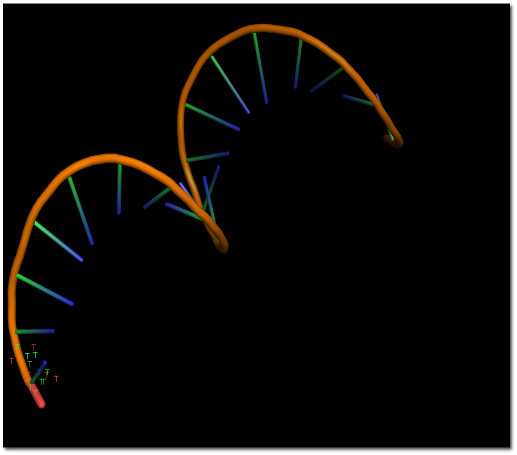
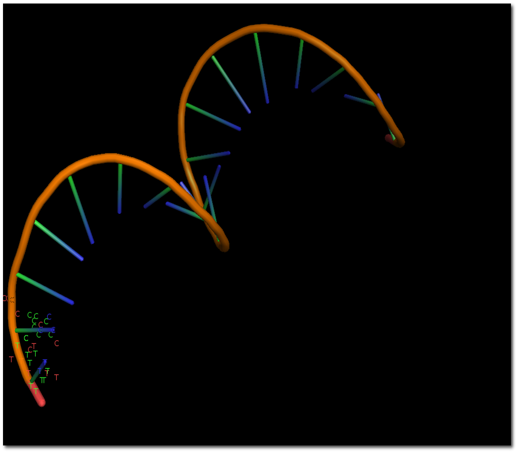
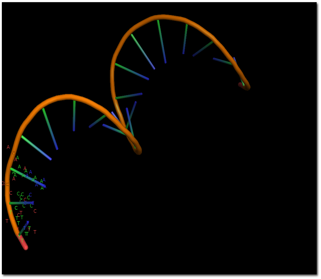
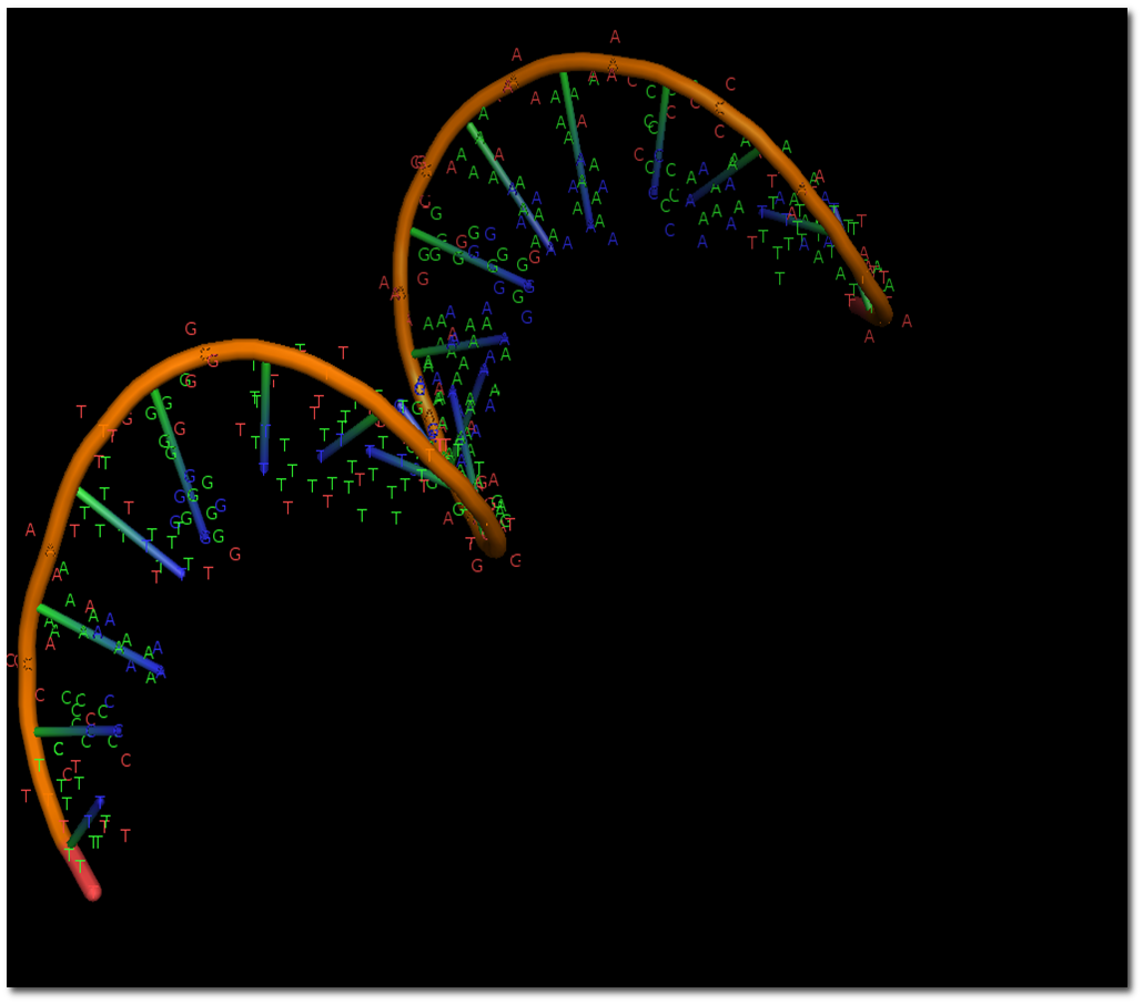
DNA double helix means DNA replication
DNA (1/15)
DNA (2/15)
DNA (3/15)
DNA (4/15)
DNA (5/15)
DNA (6/15)
DNA (7/15)
DNA (8/15)
DNA (9/15)
DNA (10/15)
DNA (11/15)
DNA (12/15)
DNA (13/15)
DNA (14/15)
DNA (15/15)
RNA structure and function
RNA contains ribose and normally uses uracil in place of thymine.
Many cellular RNAs are single strands that fold through internal base pairing.
Double-stranded RNA also exists.
Folded RNA can have structural, regulatory, or catalytic functions.
RNA secondary structure
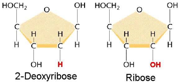
Ribose has a 2′ hydroxyl group; deoxyribose has hydrogen at that position.
RNA can form stable double helices, usually with A-form geometry. The 2′ hydroxyl affects geometry and chemical stability; it does not prevent double-stranded regions.
RNA Secondary structure
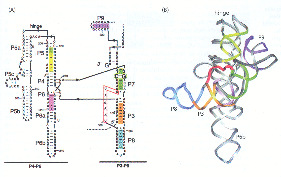
Example of three dimensional structure of RNA (Tetrahymena ribozyme)
Gene expression
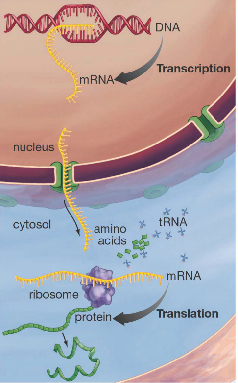
Transcription: a DNA sequence serves as a template for RNA synthesis.
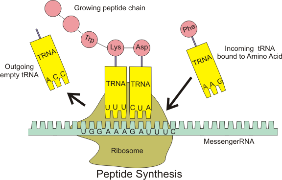
Translation: ribosomes decode an mRNA sequence to synthesise a polypeptide.
Many RNAs function without being translated.
Reverse transcription transfers information from RNA to DNA. It does not imply sequence-information transfer from protein back to nucleic acid.
The Central Dogma
The Central Dogma
Translation
The basic process of protein production is addition of one amino acid at a time to the end of a protein.
This operation is performed by a ribosome.
The ribosome molecules translate a RNA code to a specific sequence of amino acids.
tRNAs are small noncoding RNA chains (74-93 nucleotides) that transport amino acids to the ribosome.
The Central Dogma
The Central Dogma
The Central Dogma
The Central Dogma
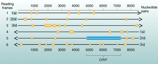
Open Reading Frames
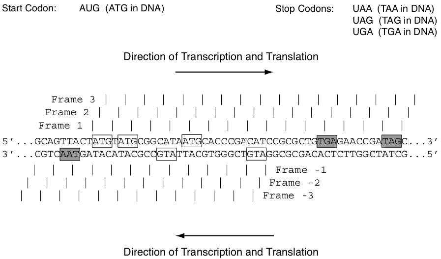
Any piece of dsDNA can be read in six possible ways, or reading frames.
For each of the two strands, either of which can be read in the 5’ to the 3’ direction.
Three possible starting points 1^{st},2^{nd},3^{rd} nuclotides.
Open Reading Frames
Open reading frames
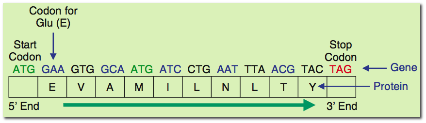
A reading frame defines how a sequence is divided into codons.
An open reading frame has no internal in-frame stop codon between its start and end under the selected genetic code.
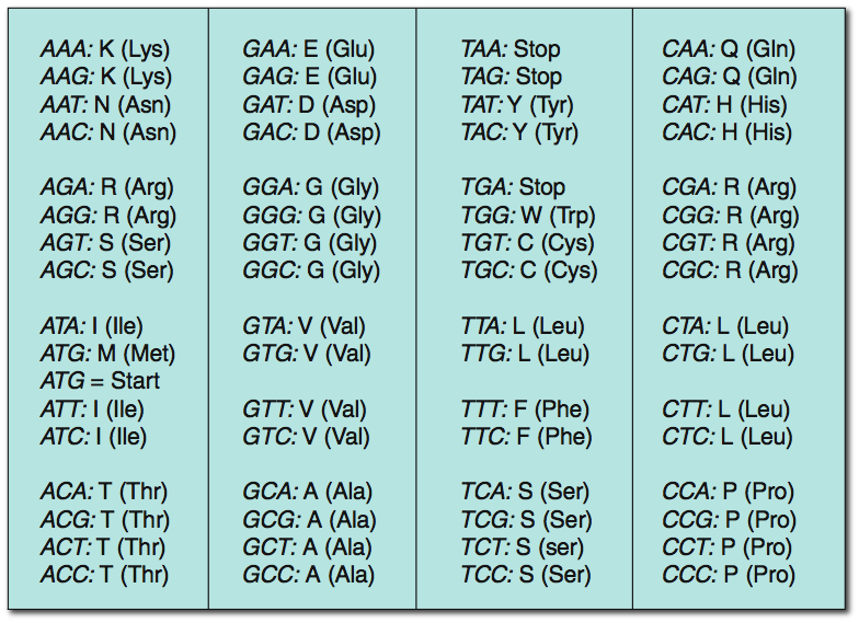
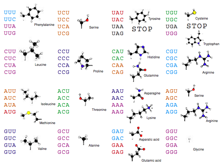
Under the standard code, UAA, UAG, and UGA are stop codons.
A long ORF is evidence for a possible coding region, but it does not prove expression or protein function.
Short ORFs can also encode proteins.
Open Reading Frames
Overlapping genes
V. Veeramachenenu Mammalian overlapping genes: the comparative perspective. Genome Res. 14:280-286,2004
DNA (1/6)
DNA (2/6)
DNA (3/6)
DNA (4/6)
DNA (5/6)
DNA (6/6)
Prokaryote vs. Eukaryote
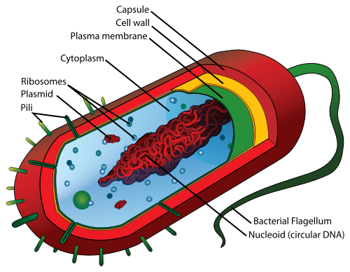
Prokaryote
pro-karyon (before kernel).
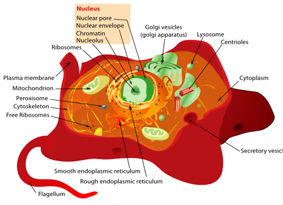
The prokaryote are a group of organisms that lack a cell nucleus , or any other membrane-bound organelles.
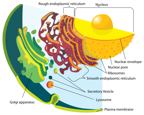
Eukaryote
eu-karyon (good kernel).
A eukaryote is an organism whose cells contain complex structures enclosed within membranes.
Prokaryote vs. Eukaryote
Prokaryote
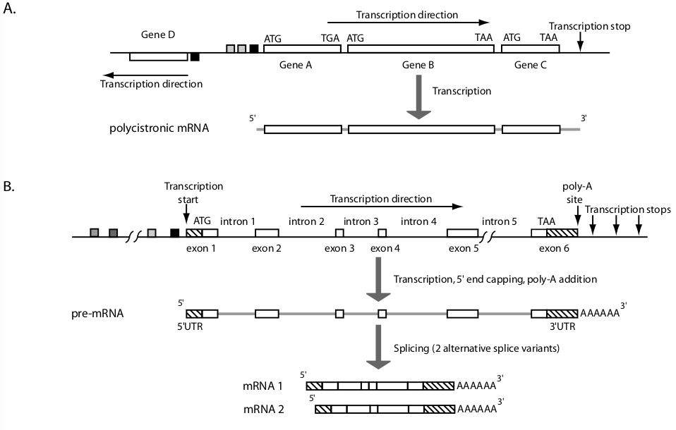
Genes in bacteria are normally close together.
May be organized as operons.
Or may be individually transcribed.
The mRNA product of the transcription is ready for immediate translation.
Eukaryote
Has transcription factor binding sites upstream of the promoter
Promoter regions may be extensive, and introns may also be long.
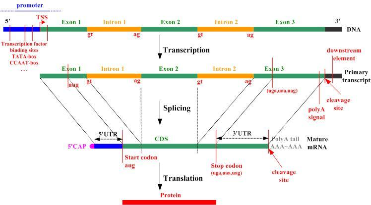
First and last exons contains regions that will not be translated (5’ UTR, 3’ UTR).
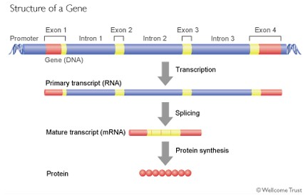
The immediate product of transcription is a pre-mRNA that is modified at the 5’ and 3’ ends.
The poly-A tail is added to the end of the message after transcription has occurred.
Splicing removes the introns to produce mature mRNA species that are ready for translation
Alternative splicing may or may not occur.
Prokaryote vs. Eukaryote
Coding sequence and ORF
A coding sequence (CDS) specifies the nucleotide sequence translated into a polypeptide.
For an intron-containing eukaryotic gene, the CDS is assembled from the coding portions of its exons.
Untranslated regions belong to the transcript but not to the CDS.
An ORF is a sequence property. A CDS is an annotation supported by biological evidence or prediction.
DNA packaging: nanometres to metres
Structure
Approximate scale
DNA double helix
2 nm wide
Nucleosome core
11 nm across
DNA around one core
147 bp, ~1.7 turns
Human cell nucleus
6 µm across*
1 µm = 1,000 nm
About 2 metres of DNA fit inside one nucleus. A typical diploid cell before DNA replication contains ~6 billion bp; 6 × 10⁹ bp × 0.34 nm/bp ≈ 2 m.
Figure: NIH / NHGRI. *Nuclear size varies by cell type. Schematic, not a fixed folding ladder: chromatin organisation is heterogeneous. The X-shaped example is a replicated chromosome with two sister chromatids.
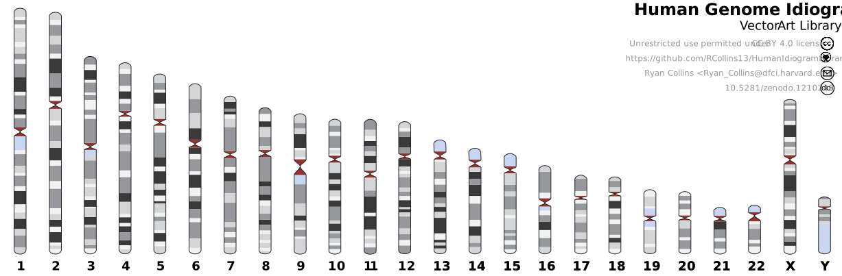
The human genome: chromosome ideogram
Chromosome complement
DNA amount
Sequence / extended DNA
23 chromosomes per typical gamete
~3 billion bp per haploid set
Chr 1: 249 Mb ≈ 8.5 cm
46 in a typical diploid cell
~6 billion bp before replication
Chr 21: 46.7 Mb ≈ 1.6 cm
24 types are shown (1–22, X, Y), not the chromosomes of one cell. Bands are cytogenetic landmarks, not individual genes. 1 Mb = 1 million bp.
Watch how the view moves from the nucleus to DNA and its molecular machinery.
WEHImovies · 7:19 · No narration; original English labels. Soundtrack retained.
Kinesin: how a molecular motor walks
Follow the two heads and the ATP → ADP cycle.
Graham Johnson · 2:09 · English subtitles: automatic transcript with terminology and segmentation corrected.
Kinesin: transporting cargo
Connect molecular stepping to transport at the scale of a cell.
em2134x · 0:22 · English viewing-guide captions (not a spoken transcript).
Your body’s molecular machines
Watch how molecular machines cooperate during cell division.
Veritasium · 6:20 · English subtitles from the published caption track. Starts at 0:02.
Case study: SARS-CoV-2, a virus written in the same code
A coronavirus is an RNA virus: its genome is a single positive-sense RNA strand of about 30,000 nucleotides — read directly by the host ribosome, no DNA step needed.
Its ~30 genes encode the components you can see in the next 3D models: spike (S), envelope (E) and membrane (M) proteins stud the lipid envelope; inside sits the nucleocapsid (N) wrapped around the RNA.
Every structural protein is specified by an open reading frame — the same reading-frame logic we just saw applies to a virus.
Sequencing this genome — and its mutations — is what made diagnostics, vaccines and variant tracking possible in weeks.
Explorable: the virion assembled from PDB structures
Rotate the virion; the protein distribution is research-based but stylised, assembled from published PDB structures of the S, E and M proteins.
Rotate, zoom and cut into the particle with the mouse or trackpad. Double-click a protein to isolate it; press the camera icons to tour the annotated parts.
Molecular binding close-up: the RBD–ACE2 interface
The receptor-binding domain (RBD) of the spike docks onto the ACE-2 peptidase domain.
The magenta receptor-binding motif (RBM) forms most of the contact — a few substitutions here (e.g. N501Y) change how tightly the virus binds, which is why mutations matter.
Despite its complexity, there exists some fundamental principles common to all cell types.
Information. e.g. Required information to create a replica of itself.
Machinery. e.g. collect materials to manufacture its own components, replicate information and boot the new cell.
Life and genetic material
Cells
Life-cycle! (born, eat, reproduce and die)
Very controlled processes
e.g. A cell only replicates if there are sufficient resources (nutrients) to complete the process
How do they decide?
Hey, they have no brain!
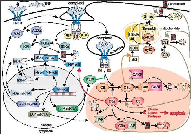
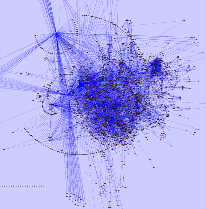
They take decisions through pathway networks.
Life and genetic material
Pathway Networks
Synthesize materials.
Break compounds.
Signal information (apoptosis).
Complex reaction networks.
Genotype and phenotype
Genotype describes an organism’s genetic constitution, either at particular loci or more broadly.
Phenotype comprises observable or measurable characteristics.
Phenotypes arise through genetic effects, environmental influences, development, and their interactions. “Genotype + environment” is a conceptual shorthand, not a universal additive equation.
Some notes on the Human Genome Project
Some notes on the Human Genome Project
Some notes on the Human Genome Project
Some notes on the Human Genome Project
Some notes on the Human Genome Project
Some notes on the Human Genome Project
Genetic variation
Mutation introduces new sequence variants; recombination reshuffles existing variation.
At a typical diploid autosomal locus, an individual carries two allele copies.
A population can have more than two alleles at the same locus.
Rare variants remain biologically relevant; a 1% frequency threshold is a historical convention for the word “polymorphism”.
Genetic Variability
In spots where no genetic variability exists, both alleles will be equal.
In spots where genetic variability exists, the alleles could be different
Homozygote: Both alleles are equal
Heterozygote: alleles are different
Mutation types: the DNA change and its consequence
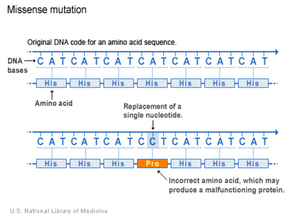
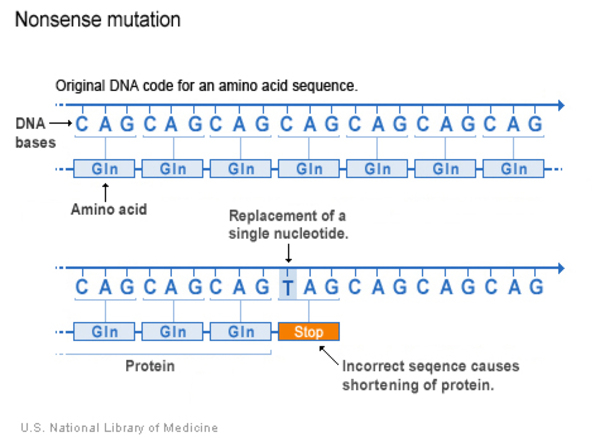
A DNA change and a protein consequence describe different things.
DNA change
Possible consequence in a coding region
Single-base substitution
Synonymous, missense or stop-gain
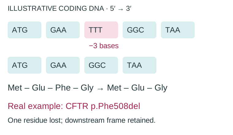
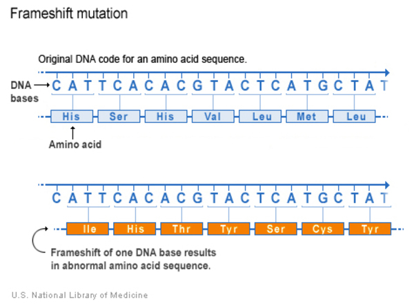
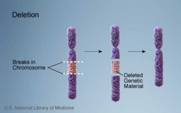
Insertion or deletion
In-frame change or frameshift
Larger rearrangement
Changes in dosage, gene structure or regulation
The consequence depends on where the change occurs and which transcript is affected.
Small DNA changes: substitution, insertion and deletion
Reference
ACGTAC
Substitute G → A
ACATAC
Insert T
ACGTTAC
Delete G
AC∅TAC
A substitution replaces a base. An insertion adds sequence. A deletion removes sequence. ∅ marks the deleted position.
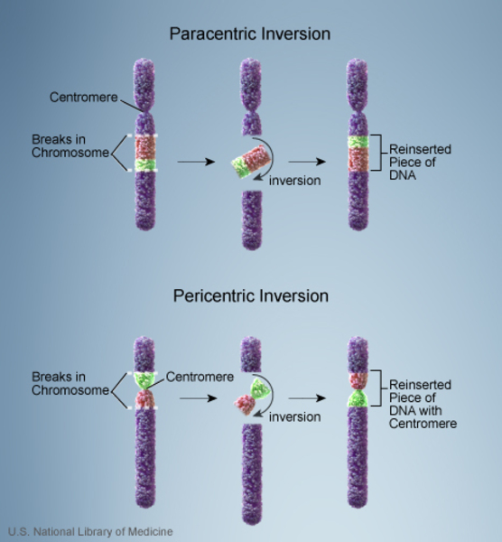
Larger changes: copy number, orientation and location
Letters below represent chromosome segments, not individual bases.
Reference
A→B→C→D→E→
Duplicate C
A→B→C→C→D→E→
Invert B–C
A→C←B←D→E→
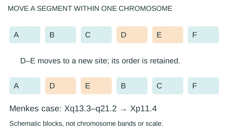
Move B–C
X→B→C→Y→
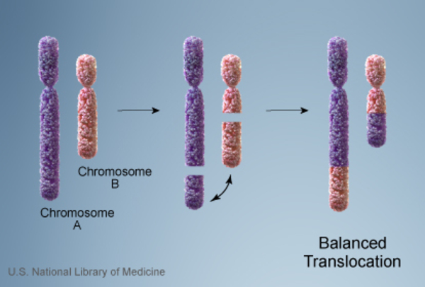
A translocation moves a segment to a new chromosomal context. An inversion reverses its orientation.
Variant names describe different properties
Term
What it describes
SNV
A difference at one nucleotide position
SNP
A single-nucleotide polymorphism in a population
Indel
An insertion or deletion of sequence
CNV
A difference in the copy number of a genomic segment
An SNV can be synonymous, missense or stop-gain in a coding region.
An indel can affect coding sequence, splicing or regulation.
Mutation types: a map from DNA change to disease
Classify what changes in DNA first; then ask what happens to RNA, protein or gene dosage.
These are teaching groups, not mutually exclusive mechanisms. Here, “point mutation” means a single-base substitution; some definitions also include one-base indels. Splicing and regulatory effects can occur across groups.
How to read mutation notation
Reference : level . position change → NM_000518.5:c.20A>T
HBB transcript/version · coding DNA · base 20 · A replaced by T
DNA: what changed?
Example¹
Read it as…
c.20A>T
Substitute A with T at base 20
c.1521_1523del
Delete bases 1521–1523 (CFTR)
c.76_77insA
Insert A between bases 76 and 77
c.76_78dup
Copy bases 76–78 next to themselves
g.100_200inv
Invert genomic bases 100–200
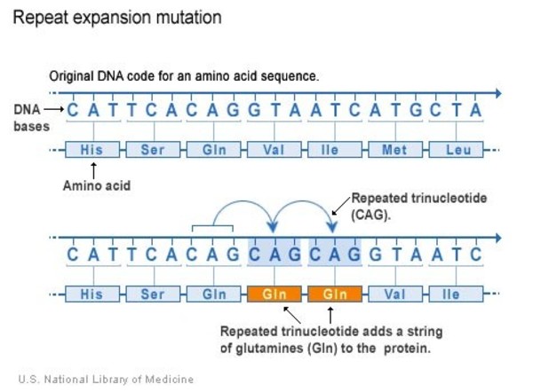
CAG[40]
40 repeats; locus/coordinates also needed
Protein: what is the consequence?
Example¹
Read it as…
p.Gly608=
Glycine unchanged: synonymous
p.Glu7Val / E7V
Glutamate → valine at residue 7
p.Gln40Ter / Q40*
Glutamine → STOP: nonsense
p.Phe508del / ΔF508
Lose phenylalanine 508: in-frame
p.Arg97ProfsTer23
Arg97 → Pro; frame shifts; STOP is codon 23 of the new frame
Levels:g. genomic DNA · c. coding DNA · r. RNA · p. protein. Chromosomes:t(9;22) means a translocation involving chromosomes 9 and 22 (cytogenetic shorthand, not HGVS).
¹ Abbreviated examples; a full HGVS report needs a reference accession and version. Parentheses mark a prediction: p.(Glu7Val). Legacy HbS E6V = HGVS p.Glu7Val because the numbering conventions differ.
The molecular change, its functional consequence, and its clinical effect are three different descriptions.
Check your classification
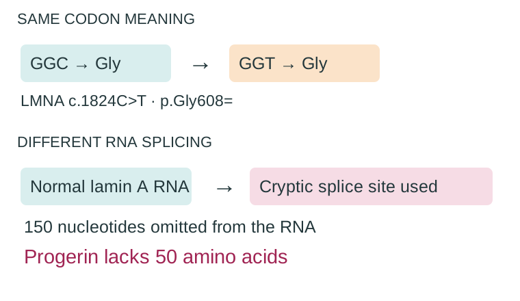
A synonymous change activates a cryptic splice site. Is it necessarily harmless?
A coding deletion removes six bases. Does it necessarily shift the reading frame?
A rearrangement has no visible loss of DNA. Can it still cause disease?
One chromosome segment moves elsewhere on that same chromosome. Is that interchromosomal?
1 No: LMNA/progeria. 2 No: six is divisible by three, assuming unchanged splicing. 3 Yes: F8 disruption or BCR::ABL1 fusion. 4 No: it is intrachromosomal relocation.